PYLARIFY AI™ enables accurate, efficient, and reproducible quantification of PYLARIFY PET/CT scans1,2
PYLARIFY AI™ is the only FDA-cleared medical device to offer standardized quantitative and accurate reporting of PSMA PET/CT images, including those achieved using PYLARIFY® PET/CT.3 Standardized reporting of PSMA assessments can improve the management of prostate cancer patients, including the accurate quantification of disease burden with increased reproducibility on a patient level.1,2
Find out how PYLARIFY AITM
can improve your workflow

Reproducible and quantitative disease-burden indices1,2
- Improved inter-reader reproducibility in staging (κ >0.80) and quantification (ICC: 0.99) of prostate cancer patients1,2
- Quantitative PSMA scan index (PSI) was associated with PSA and Gleason Score1

Accurate lesion quantification2
- High segmentation and detection accuracy (>90%) for PSMA lesions in regional and distant lymph nodes2
High efficiency1
- Significant efficiency in creating quantitative structure reporting, averaging 3-5 minutes for a comprehensive, quantitative report1
Clinical utility1
- A quantitative PSMA score/index can help stratify patients for available treatment options1
- A quantitative PSMA score/index can accurately demonstrate disease progression and response1
What is PYLARIFY AI™?2
PYLARIFY AI™ is a deep-learning application that enables2:

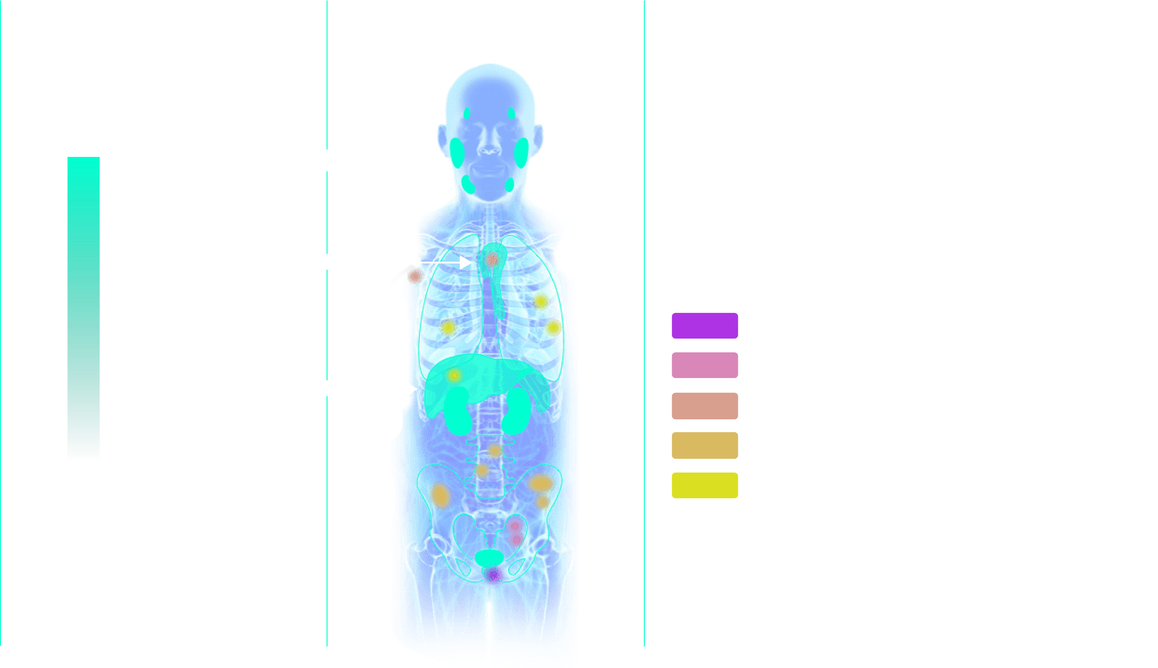
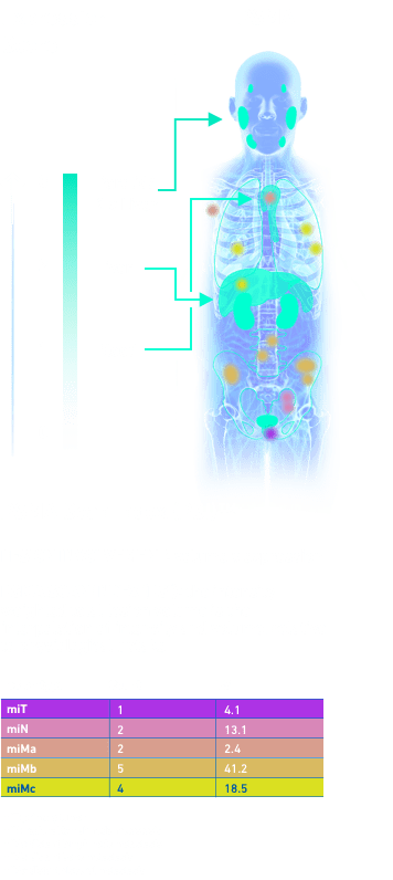
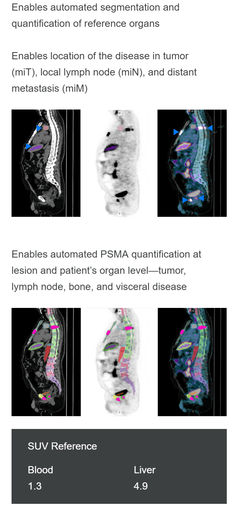
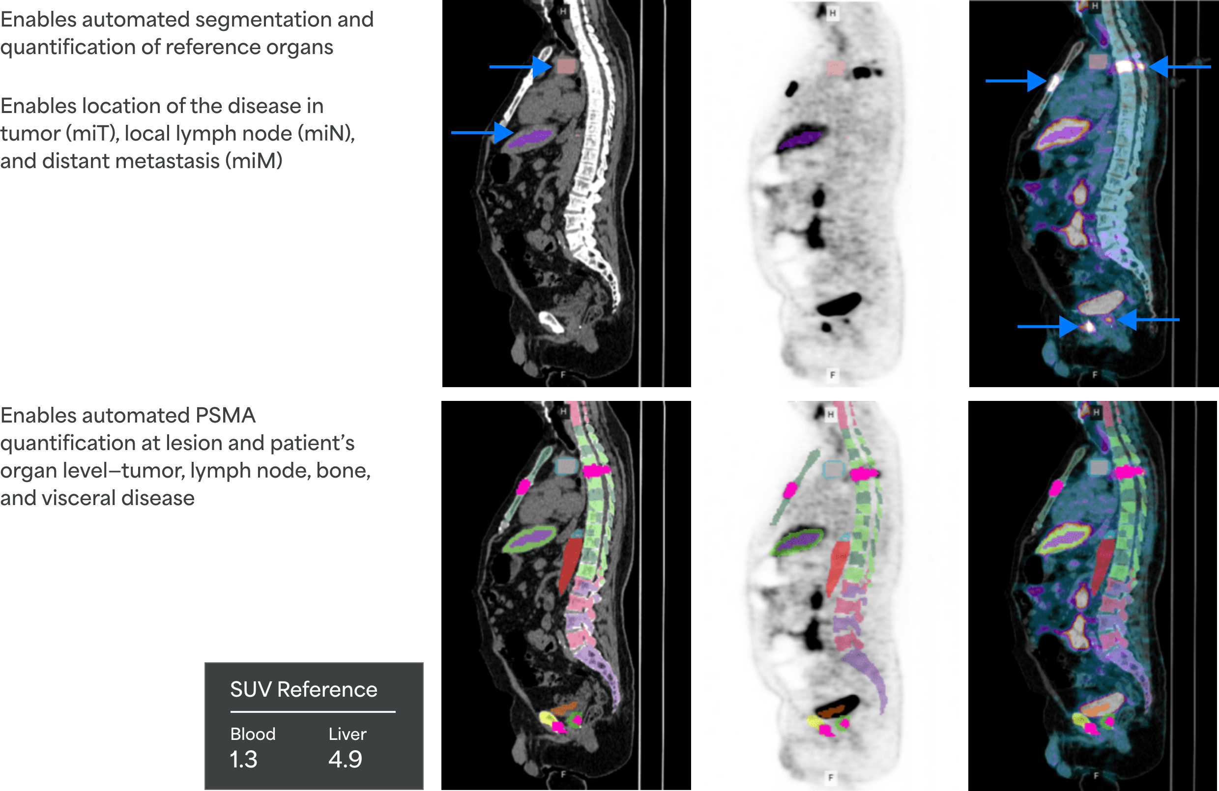
Automated segmentation and quantification of PSMA uptake in reference organs
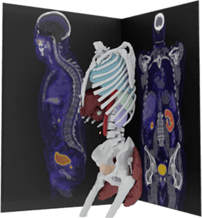
Representation and not an actual image from the software.

Localization of PSMA disease across tumor (miT), local lymph node (miN), and distant metastasis (miM)
Representation and not an actual image from the software.

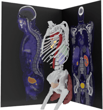
Automated PSMA quantification at lesion and organ level for patients— tumor, bone, lymph node, and visceral disease.
ITLV: Intensity-weighted Tissue Lesion Volume (ITLV) is a PSMA index
ITLV: 23.0

Representation and not an actual image from the software.

Standardized quantitative report to augment clinical impression
What does PYLARIFY AI™ report on?2,3
- PYLARIFY AI™ automatically analyzes the CT image to segment anatomical regions, including the liver and the thoracic part of the aorta, as reference organs
- Subsequently, PYLARIFY AI™ analyzes the PET image to detect target hotspots, regions (ROIs) of locally elevated PSMA tracer intensities indicative of suspicious tumor tissue and metastasis
- ROIs are marked by selecting from predefined hotspots or by manual drawing
- The selected lesions are labeled with type and location, and quantified in terms of standard uptake values (SUVs) for tracer uptake intensities and their respective volumes
- When used by radiology professionals, the standardized PYLARIFY AI™ report provides consistent and quantitative assessments of PSMA PET/CT images
Introducing the PSMA Scan Index (PSI)™1
- A unique quantitative assessment based on the unique physiological uptake of PSMA and inspired by recommended guidelines for PSMA assessment
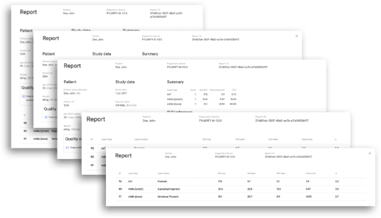
Sample report. Not an actual patient.
How does PYLARIFY AI™ work?2
Deep-learning-enabled segmentation of anatomical context in low-dose CT of PSMA PET/CT
 Sample report. Not an actual patient.
Designed to complement the clinical workflow
PYLARIFY AI™ deployment can be facilitated both as a secure web cloud application and as a local server application to secure private patient data.
Once deployed, the adaptive application can be integrated to the institution's image acquisition platforms or to the Picture Archiving and Communication Systems (PACS). The report generated from PYLARIFY AI™ is available in DICOM and JPG format and can be attached in the existing clinical impression for the physician’s review.
Seamless integration into existing clinical workflow (eg, linking to PACS) delivers a unique combination of clinical utility in a workflow-friendly package
BCR=biochemical recurrence; CI=confidence interval; CLR=correct localization rate; CT=computed tomography; MRI=magnetic resonance imaging; PCa=prostate cancer; PET=positron emission tomography; PSA=prostate-specific antigen; RP=radical prostatectomy; RT=radiation therapy.
INDICATION & IMPORTANT
SAFETY INFORMATION
PYLARIFY® (piflufolastat F 18) Injection is a radioactive diagnostic agent indicated for positron emission tomography (PET) of prostate-specific membrane antigen (PSMA) positive lesions in men with prostate cancer:
- with suspected metastasis who are candidates for initial definitive therapy.
- with suspected recurrence based on elevated serum prostate-specific antigen (PSA) level.
Radiation Risks
Diagnostic radiopharmaceuticals, including PYLARIFY, expose patients to radiation. Radiation exposure is associated with a dose-dependent increased risk of cancer. Ensure safe handling and preparation procedures to protect patients and health care workers from unintentional radiation exposure. Advise patients to hydrate before and after administration and to void frequently after administration.
INDICATION
PYLARIFY® (piflufolastat F 18) Injection is a radioactive diagnostic agent indicated for positron emission tomography (PET) of prostate-specific membrane antigen (PSMA) positive lesions in men with prostate cancer:
- with suspected metastasis who are candidates for initial definitive therapy.
- with suspected recurrence based on elevated serum prostate-specific antigen (PSA) level.
References
- Nickols N, Anand A, Johnsson K, et al. aPROMISE: a novel automated PROMISE platform to standardize evaluation of tumor burden in 18F-DCFPyL images of veterans with prostate cancer. J Nucl Med. 2022;63(2):233-239. doi:10.2967/jnumed.120.261863
- Johnsson K, Brynolfsson J, Sahlstedt H, et al. Analytical performance of aPROMISE: automated anatomic contextualization, detection, and quantification of [18F]DCFPyL (PSMA) imaging for standardized reporting. Eur J Nucl Med Mol Imaging. 2022;49(3):1041-1051. doi:10.1007/s00259-021-05497-8
- FDA clearance letter for aPROMISE X. Food and Drug Administration. April 29, 2022.