Clinical study results
OSPREY COHORT A
Patients with high-risk PCa who were candidates for initial definitive therapy
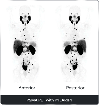
In the OSPREY study (COHORT A), PSMA-targeted PET scan with PYLARIFY significantly improved specificity over standard imaging while maintaining comparable sensitivity and delivering high PPV and NPV.1
- The co-primary endpoint for specificity was met, while the co-primary endpoint for sensitivity was not
Compared to standard imaging, PYLARIFY PET/CT delivered:
- Significantly higher specificity (97.9% vs 65.1%)
- Nearly 3 times the PPV (86.7% vs 28.3%)
- Similar sensitivity to standard imaging (40.3% vs 42.6%)
Co-primary endpoint results
- In the pre-specified analysis, the specificity co-primary endpoint with PYLARIFY® PET/CT was met (the lower limits of the 95% CIs for all readers were >80%), but the sensitivity co-primary endpoint was not met1
- PYLARIFY PET/CT achieved high PPV (range: 72%-81%) and NPV (81%-84%)2
Accurate lesion detection at initial staging vs conventional imaging¹
of patients
Patients who were thought to have localized disease based on conventional imaging were upstaged to N1 or M1 disease based on highest stage as reported by at least one reader. This upstaging of disease may have a substantial impact on treatment management decisions.*
*Future studies will be necessary to demonstrate whether PYLARIFY PET/CT-directed changes in management lead to improved outcomes for patients with prostate cancer.
OSPREY post-hoc sensitivity analysis for detection of nodal metastases >5 mm1
- Post-hoc analysis was conducted evaluating PET/CT for detection of nodal metastases >5 mm
- 27 patients were excluded whose largest nodal metastasis was ≤5 mm
- Sensitivity
- Specificity
- PPV
- NPV
Post-hoc analysis (Pelvic lymph nodes >5 mm)4 n=225
Adapted from Pienta KJ, et al. In: J Urol. 2021.
Primary analysis (All pelvic lymph nodes)2 n=252
Adapted from PYLARIFY® Prescribing Information. 2021.
CI=confidence interval; CT=computed tomography; NPV=negative predictive value; PCa=prostate cancer; PET=positron emission tomography; PPV=positive predictive value; PSA=prostate-specific antigen; PSMA=prostate-specific membrane antigen.
CONDOR
Patients with recurrent PCa based on rising PSA after therapy
In the CONDOR study, PSMA-targeted PET scan with PYLARIFY achieved the primary endpoint of correct localization rate (CLR): 85%-87% across all 3 readers (lower bound of 95% CI: 78%-80%).5 A total of 208 men with a median baseline PSA of 0.8 ng/mL (range: 0.2-98.4 ng/mL) underwent a PSMA-targeted PET scan with PYLARIFY
Correct Localization Rate (CLR) validates precision2,5
CLR is a unique endpoint that confirms lesions and pinpoints their precise location. This precision can help inform treatment decisions for your patients.
|
PYLARIFY accurately identified the existence and location of lesions with consistent findings across 3 readers |
|||
|---|---|---|---|
|
PYLARIFY accurately identified the existence and location of lesions with consistent findings across 3 readers |
Reader 1 |
Reader 2 |
Reader 3 |
|
True-positive |
89 |
87 |
84 |
|
False-positive |
15 |
13 |
15 |
|
CLR % (95% Cl) |
86 (79, 92) |
87 (80, 94) |
85 (78, 92) |
 At >80%, these results significantly exceeded the predetermined threshold of 20% in the CONDOR study5
PYLARIFY delivered high true-positives and low false-positives5
*Median result of 3 independent readers, 95% CI (78.8-92.3)
True-positives*
False-positives*
*Median result of 3 independent readers, 95% CI (78.8-92.3)
Correlative imaging5
- ¹⁸F fluciclovine PET/CT (n=71): 86.8%-90.9% across all 3 readers
- MRI (n=23): 80.0%-86.7% across all 3 readers
- CT (n=6): 80.0%-100% across all 3 readers
Histopathology5
- 78.6% to 82.8% (n=31) across all 3 readers
(36.2% at <0.5 ng/mL to 96.7% at ≥5 ng/mL)1
- Reader 1
- Reader 2
- Reader 3
95% CI
- Reader 1
- Reader 2
- Reader 3
Physicians completed pre- and post-medical management questionnaires for 205 patients
of patients
Nearly 2 out of 3 patients who underwent PSMA PET imaging with PYLARIFY after negative or uninformative standard imaging had a change in intended disease management based on findings of the PYLARIFY scan. Change in management may prevent undertreatment or overtreatment.*
Salvage local therapy
![]()
Systemic therapy
(n=58; 28.3%)
Systemic therapy
![]()
Salvage local therapy
(n=43; 21.0%)
Observation
![]()
Initiating therapy
(n=49; 23.9%)
Planned treatment
![]()
Observation
(n=9; 4.4%)
of patients with baseline PSA <0.5 ng/mL had a unidirectional change in planned management.6
*Change in treatment plan was a secondary endpoint in CONDOR. Future studies will be necessary to demonstrate whether PSMA-targeted PET scan with PYLARIFY-directed changes in management lead to improved outcomes for patients with prostate cancer.
†These data are based on responses provided by treaters on medical management questionnaires administered before the PYLARIFY scan and then 60 days after the PYLARIFY scan.
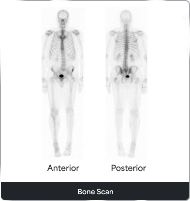
‡Standard imaging included CT/MRI/bone scan and 18F fluciclovine.1
BCR=biochemical recurrence; CI=confidence interval; CLR=correct localization rate; CT=computed tomography; MRI=magnetic resonance imaging; PCa=prostate cancer; PET=positron emission tomography; PSA=prostate-specific antigen; PSMA=prostate-specific membrane antigen.
OSPREY COHORT B
Patients with suspected recurrence or metastasis based on conventional imaging
In the OSPREY study (COHORT B), PSMA-targeted PET scan with PYLARIFY detected PSMA-positive lesions in 57.6% (19/33) of patients with no evidence of distant metastases on standard imaging, upstaging their disease and informing their treatment plan1
- 11 of 19 patients underwent targeted biopsy and 10/11 (91%) were confirmed to have prostate cancer on pathology7
- 8 of 19 patients (42%) were not able to be biopsied7
- In COHORT B (93 evaluable patients, median prostate-specific antigen 11.3 ng/mL), median sensitivity and positive predictive value for extraprostatic lesions were 95.8% (87.8%-99.0%) and 81.9% (73.7%-90.2%), respectively1
- Sensitivity
- PPV
PET=positron emission tomography; PPV=positive predictive value; PSMA=prostate-specific membrane antigen.


INDICATION
PYLARIFY (piflufolastat F 18) Injection is a radioactive diagnostic agent indicated for positron emission tomography (PET) of prostate-specific membrane antigen (PSMA) positive lesions in men with prostate cancer:
- with suspected metastasis who are candidates for initial definitive therapy.
- with suspected recurrence based on elevated serum prostate-specific antigen (PSA) level.
IMPORTANT SAFETY INFORMATION
Contraindications
None.
Warnings and Precautions
Risk of Image Misinterpretation
Imaging interpretation errors can occur with PYLARIFY imaging. A negative image does not rule out the presence of prostate cancer and a positive image does not confirm the presence of prostate cancer. The performance of PYLARIFY for imaging of patients with biochemical evidence of recurrence of prostate cancer seems to be affected by serum PSA levels. The performance of PYLARIFY for imaging of metastatic pelvic lymph nodes prior to initial definitive therapy seems to be affected by risk factors such as Gleason score and tumor stage. PYLARIFY uptake is not specific for prostate cancer and may occur with other types of cancer as well as non-malignant processes and in normal tissues. Clinical correlation, which may include histopathological evaluation of the suspected prostate cancer site, is recommended.
Hypersensitivity Reactions
Monitor patients for hypersensitivity reactions, particularly patients with a history of allergy to other drugs and foods. Reactions may be delayed. Always have trained staff and resuscitation equipment available.
Radiation Risks
Diagnostic radiopharmaceuticals, including PYLARIFY, expose patients to radiation. Radiation exposure is associated with a dose-dependent increased risk of cancer. Ensure safe handling and preparation procedures to protect patients and health care workers from unintentional radiation exposure. Advise patients to hydrate before and after administration and to void frequently after administration.
Adverse Reactions
The most frequently reported adverse reactions were headaches, dysgeusia and fatigue, occurring at rate of ≤2% during clinical studies with PYLARIFY. In addition, a delayed hypersensitivity reaction was reported in one patient (0.2%) with a history of allergic reactions.
Drug Interactions
Androgen deprivation therapy (ADT) and other therapies targeting the androgen pathway, such as androgen receptor antagonists, may result in changes in uptake of PYLARIFY in prostate cancer. The effect of these therapies on performance of PYLARIFY PET has not been established.
To report suspected adverse reactions for PYLARIFY, call 1-800-362-26681-800-362-2668 or contact FDA at 1-800-FDA-1088 or www.fda.gov/medwatch.
For important risk and use information about PYLARIFY Injection, please see Full Prescribing Information.
INDICATION
PYLARIFY (piflufolastat F 18) Injection is a radioactive diagnostic agent indicated for positron emission tomography (PET) of prostate-specific membrane antigen (PSMA) positive lesions in men with prostate cancer:
- with suspected metastasis who are candidates for initial definitive therapy.
- with suspected recurrence based on elevated serum prostate-specific antigen (PSA) level.
IMPORTANT SAFETY INFORMATION
Contraindications
None.
Warnings and Precautions
Risk of Image Misinterpretation
Imaging interpretation errors can occur with PYLARIFY imaging. A negative image does not rule out the presence of prostate cancer and a positive image does not confirm the presence of prostate cancer. The performance of PYLARIFY for imaging of patients with biochemical evidence of recurrence of prostate cancer seems to be affected by serum PSA levels. The performance of PYLARIFY for imaging of metastatic pelvic lymph nodes prior to initial definitive therapy seems to be affected by risk factors such as Gleason score and tumor stage. PYLARIFY uptake is not specific for prostate cancer and may occur with other types of cancer as well as non-malignant processes and in normal tissues. Clinical correlation, which may include histopathological evaluation of the suspected prostate cancer site, is recommended.
Hypersensitivity Reactions
Monitor patients for hypersensitivity reactions, particularly patients with a history of allergy to other drugs and foods. Reactions may be delayed. Always have trained staff and resuscitation equipment available.
Radiation Risks
Diagnostic radiopharmaceuticals, including PYLARIFY, expose patients to radiation. Radiation exposure is associated with a dose-dependent increased risk of cancer. Ensure safe handling and preparation procedures to protect patients and health care workers from unintentional radiation exposure. Advise patients to hydrate before and after administration and to void frequently after administration.
Adverse Reactions
The most frequently reported adverse reactions were headaches, dysgeusia and fatigue, occurring at rate of ≤2% during clinical studies with PYLARIFY. In addition, a delayed hypersensitivity reaction was reported in one patient (0.2%) with a history of allergic reactions.
Drug Interactions
Androgen deprivation therapy (ADT) and other therapies targeting the androgen pathway, such as androgen receptor antagonists, may result in changes in uptake of PYLARIFY in prostate cancer. The effect of these therapies on performance of PYLARIFY PET has not been established.
To report suspected adverse reactions for PYLARIFY, call 1-800-362-26681-800-362-2668 or contact FDA at 1-800-FDA-1088 or www.fda.gov/medwatch.
For important risk and use information about PYLARIFY Injection, please see Full Prescribing Information.
References
- Pienta KJ, Gorin MA, Rowe SP, et al. A phase 2/3 prospective multicenter study of the diagnostic accuracy of prostate specific membrane antigen PET/CT with 18F-DCFPyL in prostate cancer patients (OSPREY). J Urol. 2021;206(1):52-61.
- PYLARIFY® [package insert]. North Billerica, MA: Progenics Pharmaceuticals, Inc., a Lantheus company.
- Carroll PR, Probst S, Rowe SP, et al. Changes to initial risk assessment and intended patient management in high-risk prostate cancer: an exploratory analysis of cohort A from the OSPREY trial. J Urol. 2021;206(suppl 3):e181-e182.
- Pienta KJ, Gorin MA, Rowe SP, et al. A phase 2/3 prospective multicenter study of the diagnostic accuracy of prostate specific membrane antigen PET/CT with 18F-DCFPyL in prostate cancer patients (OSPREY). Supplementary Appendix. J Urol. 2021;206(1):52-61.
- Morris MJ, Rowe SP, Gorin MA, et al. Diagnostic performance of 18F-DCFPyL-PET/CT in men with biochemically recurrent prostate cancer: results from the CONDOR phase III, multicenter study. Clin Cancer Res. 2021;27(13):3674-3682.
- Pouliot F, Gorin MA, Rowe SP, et al. Changes in planned disease management after piflufolastat F 18 PET/CT in men with biochemically recurrent prostate cancer and low PSA levels: A secondary analysis of results from the CONDOR study. Poster presented at: 2023 ASCO Genitourinary Cancers Symposium; February 16-18. Abstract ID: 61.
- Data on file. Bedford, MA: Progenics Pharmaceuticals, Inc.; 2024.